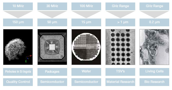
The unique characteristic of acoustic microscopy is in the ability to image the interaction of acoustic waves with the elastic properties of a specimen with the resolution of optical light microscopy. In this way the microscope is used to image the interior of an opaque material. Lower frequencies, 5 to 100 MHz. are used to achieve greater penetration in substrates. Such applications often include examination of packaging architecture to ensure integrity, and the presence of defects.
For more rigid materials, including most metals, semiconductors and ceramics, surface Raleigh waves can play a dominant role in the image contrast Propagation of the Rayleigh waves are sensitive to any disturbance in regularity within the surface structure. If the specimen is anisotropic, then there will be dependence on the orientation of the surface and the direction of propagation in it.
If there are surface cracks or boundaries, there will be a strong contrast as they scatter the Rayleigh waves. To take advantage of these acoustic phenomena frequencies in the range 800-2000 MHz are required.
In summary, low transducer frequencies provide high penetration depth but little image resolution. Higher frequencies give rise to improved image resolution while compromising penetration depth. This factor must be taken into consideration during analysis depending on the goal of the investigation. PVA TePla Analytical Systems works closely with customers to choose the correct transducer parameter set and system configuration that best suits their needs.
